Inauguration of the Latest CUVIS, the World’s Most Advanced Fully Active Robotic Joint Replacement System
by IT and Industries Minister Shri D. Sridhar Babu
An electrocardiogram, commonly called an ECG or EKG, is the most widely used cardiac investigation in medicine. It records the electrical activity of the heart through electrodes placed on the skin of the chest, arms, and legs, producing a trace that cardiologists use to evaluate heart rate, rhythm, conduction, and to identify evidence of heart attack, ischemia, hypertrophy, and many other cardiac conditions.
At Germanten Hospital, Attapur, a resting 12-lead ECG is available on a walk-in basis with same-day reporting. All ECGs are reviewed and interpreted by our senior cardiologist team. Results are discussed with the patient at the time of reporting and integrated into the wider clinical picture if further investigation or treatment is needed.
With each heartbeat, the heart generates a wave of electrical activity that spreads from the sinoatrial node in the right atrium, through the atrial muscle, down through the AV node and the bundle of His, and out through the Purkinje fibers into the ventricular muscle. A 12-lead ECG records this electrical wave from 12 different angles simultaneously, providing a comprehensive view of the heart's electrical behavior from every direction.
Represents atrial depolarization, the electrical activation of the upper chambers. Abnormal P waves indicate atrial enlargement, rhythm disturbances, or conduction delay between atria.
The time from the start of atrial activation to the start of ventricular activation, reflecting conduction through the AV node. Prolonged PR interval indicates first-degree heart block.
Represents ventricular depolarization, the electrical activation of the lower chambers. Width, morphology, and axis of the QRS reveal bundle branch blocks, ventricular hypertrophy, and prior heart attack.
The segment between ventricular activation and repolarization. Elevation indicates acute myocardial infarction or pericarditis. Depression indicates ischemia or posterior infarction. Critical for heart attack diagnosis.
Represents ventricular repolarization. Inversion can indicate ischemia, hypertrophy, or cardiomyopathy. Peaked T waves can indicate hyperkalemia (dangerously high potassium).
The total time for ventricular activation and recovery. Prolonged QT interval increases risk of dangerous ventricular arrhythmias including Torsades de Pointes. Measured to assess medication safety and congenital long QT syndrome.
Calculated from the interval between consecutive QRS complexes. Normal resting rate is 60 to 100 beats per minute.
Whether the rhythm is regular or irregular, and whether it originates from the sinus node (sinus rhythm) or from an abnormal site (ectopic rhythm). Atrial fibrillation, flutter, SVT, and ventricular arrhythmias all produce characteristic ECG patterns.
ST elevation on ECG is the key finding that triggers emergency angioplasty. Early ECG in chest pain is time-critical. Every minute of delay matters.
Q waves and ST-T changes from a previous heart attack persist on ECG for life, allowing identification of prior cardiac events even decades later.
ST depression and T-wave changes during chest pain or on a resting ECG indicate reduced blood supply to the heart muscle.
The hallmark ECG finding is absence of P waves and an irregularly irregular ventricular rhythm. Identified immediately on a 12-lead ECG if present at the time of recording.
Prolonged PR interval, dropped beats, or complete dissociation between P waves and QRS complexes reveal first, second, or third-degree AV block.
Abnormal QRS morphology indicating delayed conduction down the left or right bundle of His, which can be a sign of underlying cardiac disease or a marker of increased cardiac risk.
Increased QRS voltage indicating thickening of the left ventricle, commonly from hypertension or hypertrophic cardiomyopathy.
A short PR interval and delta wave (slurred upstroke of the QRS) indicate an accessory pathway capable of causing SVT and, in some cases, sudden cardiac death during atrial fibrillation.
A characteristic right precordial ST pattern associated with risk of sudden cardiac death from ventricular fibrillation, often in young adults without structural heart disease.
Prolonged QT interval indicating risk of Torsades de Pointes ventricular arrhythmia. May be congenital or caused by medications.
Diffuse saddle-shaped ST elevation across multiple leads, typically with PR depression, distinguishable from the localized ST elevation of heart attack.
Peaked T waves in hyperkalemia, flattened T waves in hypokalemia, and prolonged QT in hypocalcemia all have characteristic ECG patterns that can guide urgent management.
No special preparation is required for a resting ECG. You can eat and drink normally beforehand. Wear clothing that allows easy access to the chest, arms, and lower legs.
Ten electrodes (small adhesive patches with gel) are placed on specific positions on your chest, both wrists, and both ankles. The positions are standardized internationally.
You lie still and breathe normally for approximately 10 seconds while the ECG machine records electrical signals from all 12 leads simultaneously. Movement or deep breathing can introduce artifact into the trace.
The entire procedure takes 5 to 10 minutes from electrode placement to completed recording.
The ECG is entirely painless. No electricity is passed into your body. The machine only records signals generated by your own heart.
At Germanten, all ECG tracings are reviewed by a senior cardiologist on the same day. The report is discussed with you directly and any abnormal findings are acted upon immediately.
An ECG is a snapshot of the heart's electrical activity at one moment in time. A normal resting ECG does not rule out all cardiac conditions. Intermittent arrhythmias, for example, may not be present during the recording. If symptoms are intermittent, a Holter monitor or stress test may be needed in addition.
An ECG records electrical activity. A 2D echo records mechanical function, chamber size, valve anatomy, and wall motion. They provide complementary information. An ECG can detect a prior heart attack; an echo shows how much function was lost.
A resting ECG captures 10 seconds of rhythm. A Holter monitor records continuously for 24 to 48 hours, capturing intermittent arrhythmias missed on a standard ECG.
A resting ECG shows the heart at rest. A treadmill stress test records the ECG during exercise, revealing ischemic changes that only appear when the heart is working hard and blood supply is challenged.
An ECG detects electrical consequences of coronary disease (ischemia, prior infarction). CT angiography directly images the coronary arteries to visualize plaque and blockages before electrical or functional changes occur.
Consult expert cardiologists in Hyderabad at Germanten Hospital

I visited Germanten Hospital for chest pain. The cardiologist explained my heart condition clearly, reviewed my ECG, and guided me with the right treatment plan. The staff was incredibly helpful.
Germanten is a trusted hospital for heart care. My tests went smoothly, and the doctor explained the reports in simple language. I felt completely comfortable throughout the visit.
I consulted their heart specialist for high blood pressure. The doctor listened patiently and gave practical advice for managing my heart health. Excellent patient care.
My father needed guidance for a heart angiography. The cardiology team here is fantastic—they are some of the best doctors we have met. They explained every step of the procedure clearly.
I had a great experience with the cardiology department. From booking the appointment to the consultation, everything was managed perfectly. The doctor gave honest and clear advice
I went to Germanten for a preventive heart check-up. The specialist evaluated my risk factors and suggested simple, effective lifestyle changes. It was a very reassuring visit.


Watch our cardiologists explain common heart symptoms, risk factors, diagnostic tests and preventive measures.

Expert advice on maintaining cardiovascular health
Preventive measures for cardiac conditions

Understanding common heart warning signs

Overview of cardiac diagnostic procedures
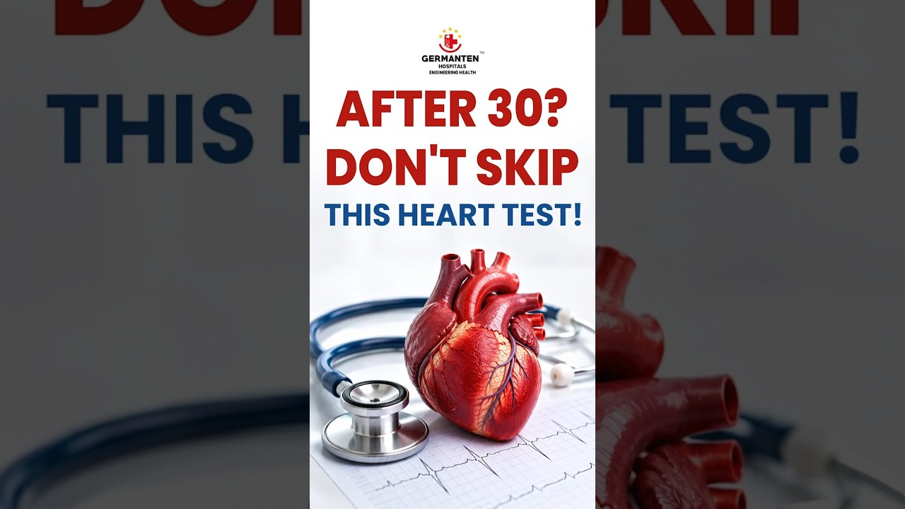
Key risk factors for cardiovascular disease
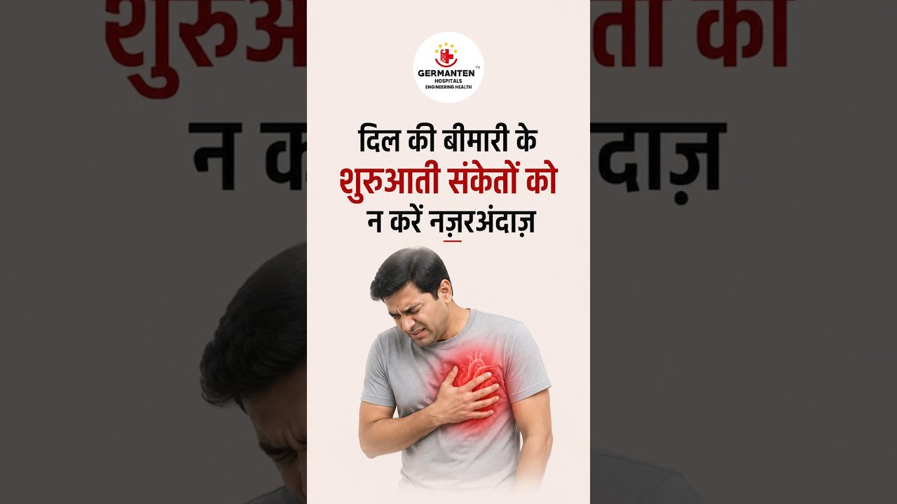
Modern treatment approaches for heart conditions
by IT and Industries Minister Shri D. Sridhar Babu
by Health Minister Shri Damodar Rajanarsimha
 01 / 12
Connect with our experts doctors for guidance
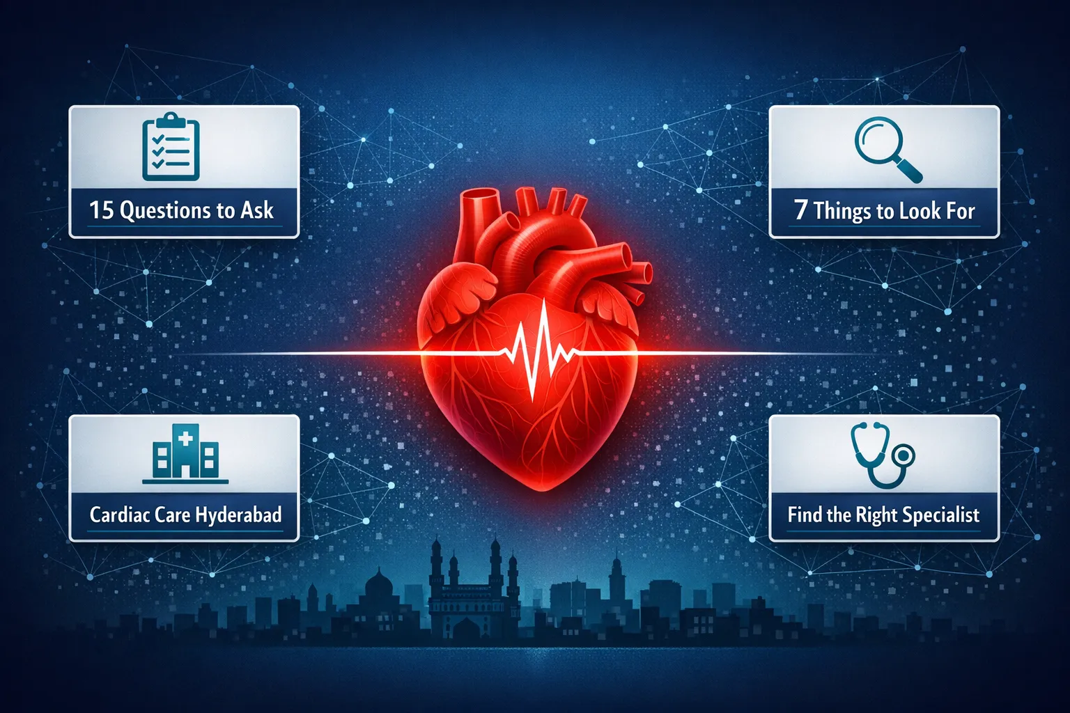
Find the best cardiologist in Hyderabad. Know what to look for, red flags to avoid, and 15 must-ask questions before your first cardiac specialist visit
Read MoreLearn how a cardiologist supports better heart health through diagnosis, prevention, regular check-ups, lifestyle guidance and cardiac care in Hyderabad
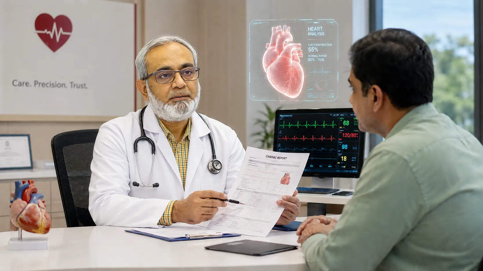
Read MoreLooking for the best cardiologist in Attapur Hyderabad? Learn what to look for, key credentials to check, and why Dr Mohammed Wasif Azam Germanten at Germanten Hospital is a trusted choice for heart care
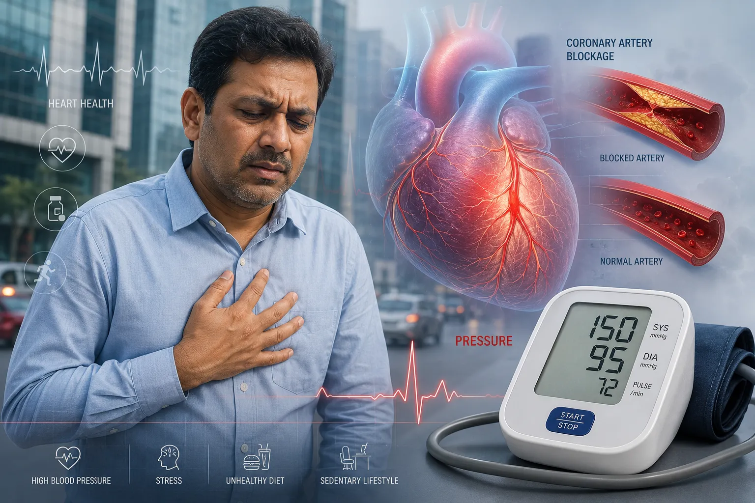
Read MoreUnderstand how high blood pressure silently damages your heart. A Hyderabad cardiologist explains the link between hypertension and heart disease, risk factors, and treatment options for patients in Attapur and across Hyderabad.
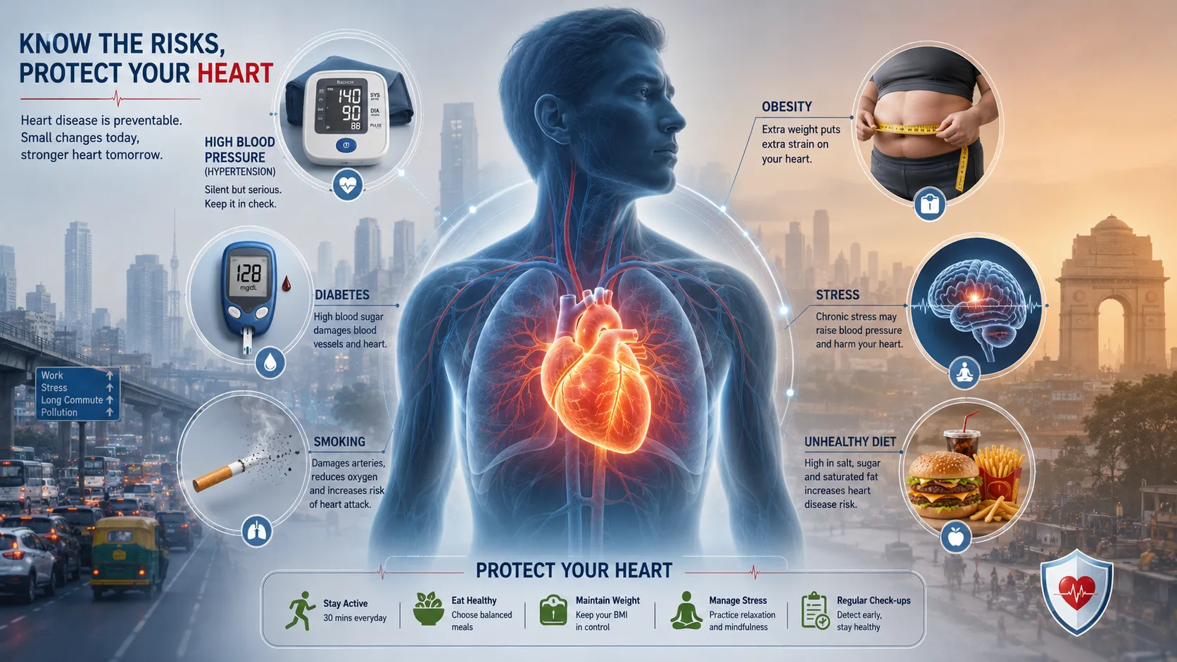
Read MoreDiscover the 10 most common heart disease risk factors in Hyderabad and how to reduce them. Expert advice from a preventive cardiologist in Attapur on diet, lifestyle, and cardiovascular prevention.
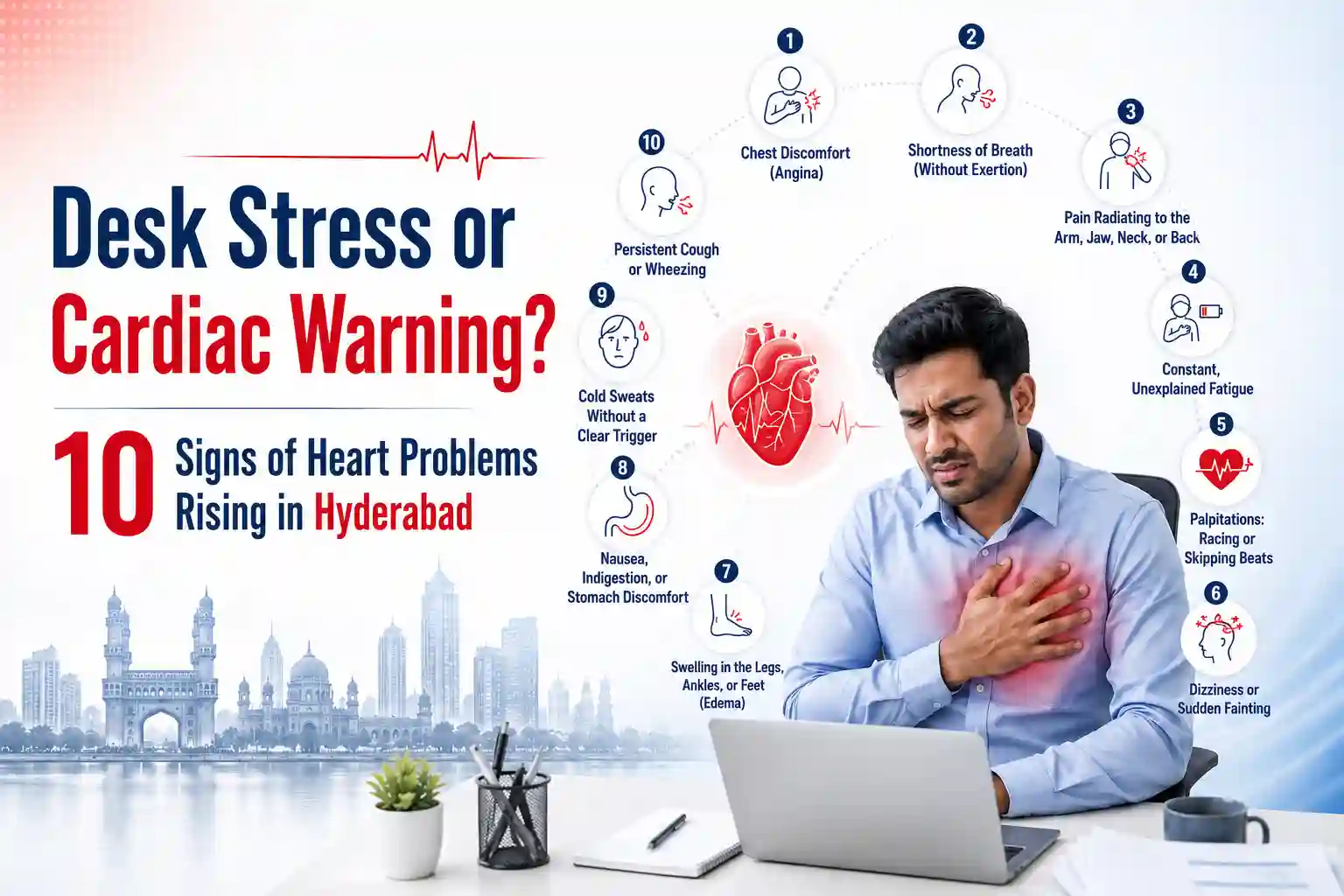
Read MoreExperiencing chest tightness or desk fatigue? Learn the 10 critical heart problem symptoms corporate workers in Hyderabad ignore. Book a checkup today.
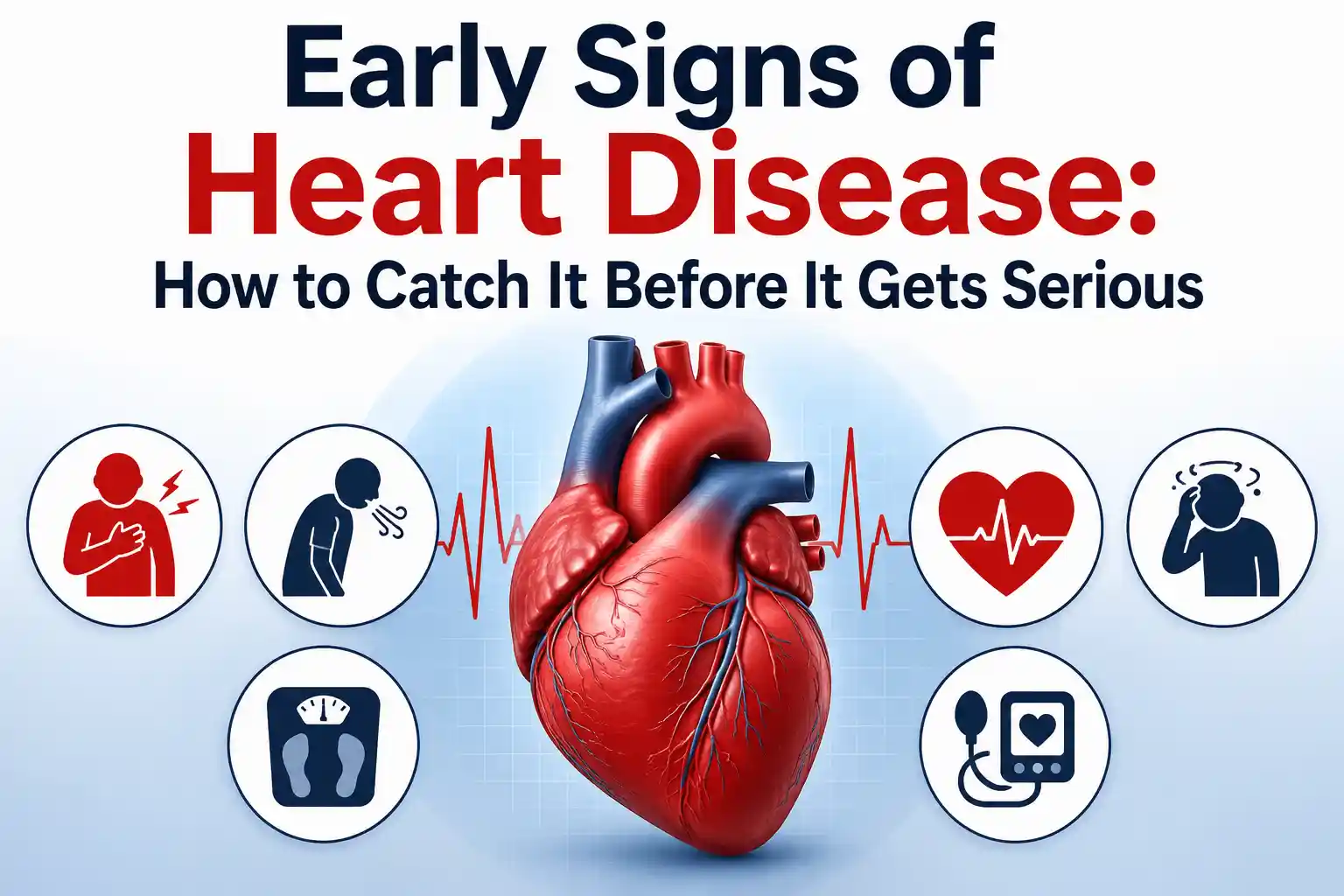
Read MoreSpot the early signs of heart disease early, from chest pain to silent symptoms. Get expert cardiology care at Germanten Hospitals, Attapur, Hyderabad.
Read MoreGermanten Hospital is located in Attapur, one of South Hyderabad's most accessible neighborhoods. Patients from across southern and western Hyderabad can reach us within 20 to 30 minutes.