Inauguration of the Latest CUVIS, the World’s Most Advanced Fully Active Robotic Joint Replacement System
by IT and Industries Minister Shri D. Sridhar Babu
TAVI, or transcatheter aortic valve implantation (also called TAVR, transcatheter aortic valve replacement), is a minimally invasive procedure that replaces a diseased aortic valve without open-heart surgery. Using a catheter guided through the femoral artery in the groin, a new biological valve is delivered to the heart and deployed precisely within the diseased valve, restoring normal function immediately.
TAVI was originally developed for patients too high-risk for conventional open-heart surgery. A decade of clinical trials has since demonstrated outcomes equivalent or superior to surgical valve replacement across a broadening range of patients. Today, TAVI is a mainstream treatment for severe aortic stenosis and is increasingly performed in intermediate and lower-risk patients at experienced structural heart centers.
The aortic valve sits between the left ventricle and the aorta, the body's main artery. It opens to allow blood to be pumped out of the heart and closes to prevent backflow. In aortic stenosis, the valve leaflets become thickened and calcified over time, reducing the valve opening and restricting blood flow out of the heart.
The heart compensates initially by pumping harder, causing the left ventricle to thicken. As the valve becomes severely narrowed, the heart can no longer compensate. The classic symptoms of severe aortic stenosis, chest pain on exertion, fainting or near-fainting, and breathlessness, signal a point at which the condition becomes rapidly life-threatening without valve replacement. Without treatment, the median survival after symptom onset is 2 to 3 years.
Aortic stenosis is the most common valve disease requiring intervention in adults over 65, and its prevalence rises sharply with age. It affects approximately 3 to 5% of people over 75 in developed populations.
Patients with severe aortic stenosis who are considered too high-risk for conventional open-heart surgery due to advanced age, frailty, previous cardiac surgery, significant lung disease, or major comorbidities. TAVI was originally validated in this group.
Multiple large trials (PARTNER 2, SURTAVI) demonstrated that TAVI is non-inferior to surgical valve replacement in intermediate-risk patients, with lower rates of stroke and bleeding.
The PARTNER 3 and Evolut Low Risk trials showed TAVI superiority or equivalence to surgery in low-risk patients under 70 at two-year follow-up, leading to expanding use in younger patients.
Patients with a previously implanted surgical bioprosthetic valve that has deteriorated can have a TAVI valve deployed within the old valve without reoperation.
TAVI requires adequate femoral artery size for catheter access and appropriate aortic anatomy. CT assessment determines suitability before the procedure.
Patients suitable for both TAVI and surgical valve replacement are discussed at our structural heart multidisciplinary team meeting, which includes interventional cardiologists and cardiac surgeons. The goal is always the option most likely to provide the best long-term outcome for that individual. Learn more about surgical valve replacement.
A CT scan of the aorta and access vessels is essential for planning. It measures the size of the aortic valve annulus to select the correct prosthesis size and assesses the access route. A coronary angiogram is also performed to evaluate the coronary arteries before proceeding.
The heart team formally agrees on the procedure based on imaging, risk assessment, and patient preferences.
TAVI is performed under general anesthesia or, increasingly, under conscious sedation (the patient is sedated but breathing independently). Sedation is preferred at many centers for lower-risk patients as it reduces intensive care admission and hospital stay.
In the transfemoral approach (preferred in over 85% of cases), a sheath is placed in the femoral artery in the groin. Alternative access sites (subclavian artery, transaortic, or transcaval) are used when the femoral vessels are too small or diseased.
A guidewire is passed through the diseased aortic valve under X-ray imaging.
In some cases, a balloon is first inflated within the narrowed valve to create space for the prosthesis. This step is omitted with some newer valve systems.
The replacement valve, a biological valve made from bovine or porcine pericardium mounted on a metal frame, is delivered on a catheter and positioned precisely at the level of the native aortic valve. The valve is then expanded, either by balloon inflation or self-expansion, and immediately begins functioning.
Echocardiography and angiography confirm correct valve positioning, function, and the absence of significant leakage.
The access site is closed with a vascular closure device. Most patients are transferred to the cardiac ward (not ICU) the same day and are discharged within 2 to 3 days.
The TAVI procedure typically takes 1 to 2 hours. Most patients are walking the following morning and discharged within 2 to 3 days, compared to 5 to 7 days for surgical valve replacement.
TAVI: Catheter through a small groin puncture. Surgery: Full sternotomy (breastbone divided).
TAVI: Not required. Surgery: Required for conventional open repair.
TAVI: 2 to 3 days. Surgery: 5 to 8 days.
TAVI: 1 to 2 weeks to light activity. Surgery: 6 to 8 weeks.
TAVI: Slightly higher in some trials. Newer devices and embolic protection have reduced this significantly.
TAVI: Slightly higher rate of mild paravalvular leak. Rarely clinically significant with modern devices.
Surgical valves have 20+ year follow-up data. TAVI valves have 10-year data showing excellent durability; longer-term data accumulating.
TAVI: Higher pacemaker requirement (10 to 20%) due to conduction system disruption. Surgery: Lower (2 to 5%).
Consult expert cardiologists in Hyderabad at Germanten Hospital

I visited Germanten Hospital for chest pain. The cardiologist explained my heart condition clearly, reviewed my ECG, and guided me with the right treatment plan. The staff was incredibly helpful.
Germanten is a trusted hospital for heart care. My tests went smoothly, and the doctor explained the reports in simple language. I felt completely comfortable throughout the visit.
I consulted their heart specialist for high blood pressure. The doctor listened patiently and gave practical advice for managing my heart health. Excellent patient care.
My father needed guidance for a heart angiography. The cardiology team here is fantastic—they are some of the best doctors we have met. They explained every step of the procedure clearly.
I had a great experience with the cardiology department. From booking the appointment to the consultation, everything was managed perfectly. The doctor gave honest and clear advice
I went to Germanten for a preventive heart check-up. The specialist evaluated my risk factors and suggested simple, effective lifestyle changes. It was a very reassuring visit.


Watch our cardiologists explain common heart symptoms, risk factors, diagnostic tests and preventive measures.

Expert advice on maintaining cardiovascular health

Preventive measures for cardiac conditions

Understanding common heart warning signs

Overview of cardiac diagnostic procedures

Key risk factors for cardiovascular disease
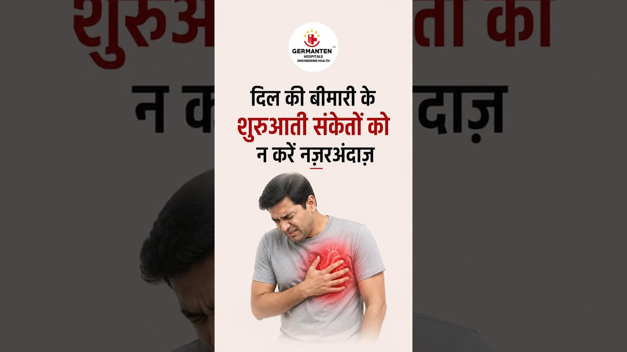
Modern treatment approaches for heart conditions
by IT and Industries Minister Shri D. Sridhar Babu
by Health Minister Shri Damodar Rajanarsimha
 01 / 12
Connect with our experts doctors for guidance
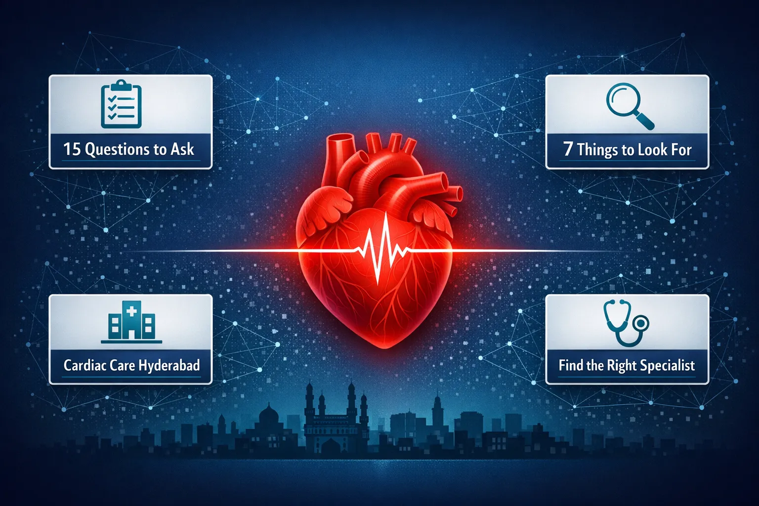
Find the best cardiologist in Hyderabad. Know what to look for, red flags to avoid, and 15 must-ask questions before your first cardiac specialist visit
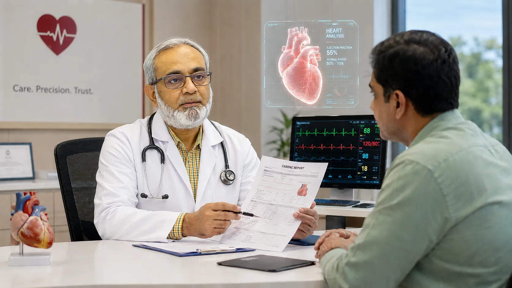
Read MoreLearn how a cardiologist supports better heart health through diagnosis, prevention, regular check-ups, lifestyle guidance and cardiac care in Hyderabad
Read MoreLooking for the best cardiologist in Attapur Hyderabad? Learn what to look for, key credentials to check, and why Dr Mohammed Wasif Azam Germanten at Germanten Hospital is a trusted choice for heart care
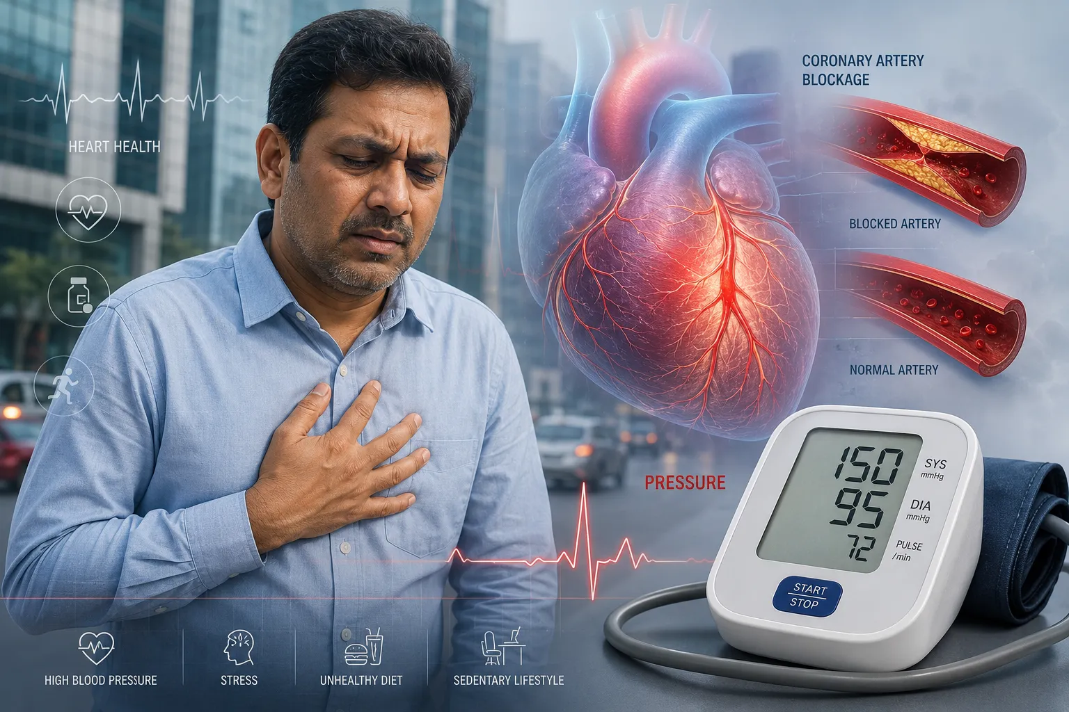
Read MoreUnderstand how high blood pressure silently damages your heart. A Hyderabad cardiologist explains the link between hypertension and heart disease, risk factors, and treatment options for patients in Attapur and across Hyderabad.
Read MoreDiscover the 10 most common heart disease risk factors in Hyderabad and how to reduce them. Expert advice from a preventive cardiologist in Attapur on diet, lifestyle, and cardiovascular prevention.
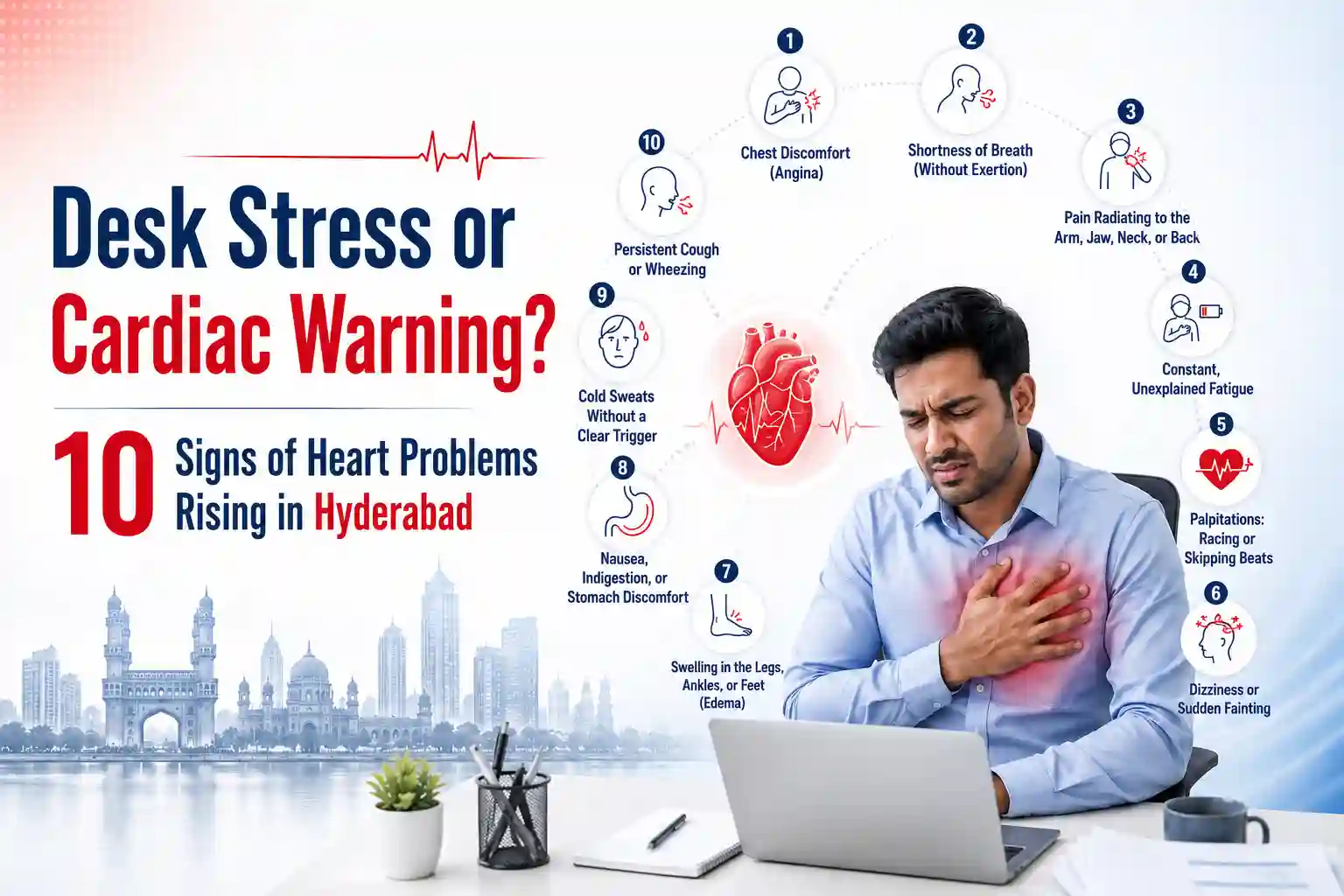
Read MoreExperiencing chest tightness or desk fatigue? Learn the 10 critical heart problem symptoms corporate workers in Hyderabad ignore. Book a checkup today.
Read MoreSpot the early signs of heart disease early, from chest pain to silent symptoms. Get expert cardiology care at Germanten Hospitals, Attapur, Hyderabad.
Read MoreGermanten Hospital is located in Attapur, one of South Hyderabad's most accessible neighborhoods. Patients from across southern and western Hyderabad can reach us within 20 to 30 minutes.