Inauguration of the Latest CUVIS, the World’s Most Advanced Fully Active Robotic Joint Replacement System
by IT and Industries Minister Shri D. Sridhar Babu
A pacemaker is a small electronic device implanted under the skin near the collarbone that monitors the heart's rhythm and delivers gentle electrical impulses to maintain an adequate heart rate when the heart beats too slowly or pauses unexpectedly. Modern pacemakers are remarkably sophisticated, adapting their pacing rate to the patient's activity level, monitoring heart rhythm continuously, and storing data that cardiologists can review remotely or at clinic visits.
Pacemaker implantation at Germanten Hospital is performed by our electrophysiology team as a minimally invasive procedure under local anesthesia. Most patients are discharged the following day and return to normal daily activities within one to two weeks. Over one million pacemakers are implanted worldwide each year, and the procedure has an excellent safety record when performed by experienced operators.
A pacemaker is indicated when the heart's natural electrical system fails to maintain an adequate rate or rhythm. The most common reasons for pacemaker implantation include:
A heart rate that is persistently too slow (typically below 40 to 50 beats per minute) causing symptoms of dizziness, fainting, breathlessness, or extreme fatigue. The most common indication for pacemaker implantation.
The sinus node (the heart's natural pacemaker) fails to fire reliably, causing the heart rate to fluctuate between very slow and fast, often associated with pauses.
A disruption in the electrical conduction pathway between the upper and lower chambers. Second-degree Mobitz type II and complete (third-degree) heart block require pacemaker implantation even in the absence of symptoms, due to the risk of sudden cardiac arrest.
Some patients with atrial fibrillation have an excessively slow ventricular response, particularly when rate-controlling medications are added. A pacemaker ensures an adequate minimum rate.
Procedures that affect the conduction system, including TAVI, some valve surgeries, and atrioventricular nodal ablation for rate control in AFib, may require pacemaker support.
An exaggerated slowing of the heart rate in response to neck pressure (turning the head, tight collars) causing recurrent fainting.
One lead placed in the right ventricle. Used when only ventricular pacing is required and atrial sensing is not needed. Simpler and less expensive than dual-chamber devices.
Two leads: one in the right atrium and one in the right ventricle. Senses the atrium and coordinates pacing of the ventricle to maintain the natural sequence of heart contraction. More physiological than single-chamber pacing and the preferred option for most patients.
Contains an accelerometer or minute ventilation sensor that detects physical activity and increases pacing rate accordingly, allowing patients to exercise normally. Most modern devices are rate-responsive.
A specialized device with three leads: right atrium, right ventricle, and left ventricle. Used in patients with heart failure and electrical conduction delay (broad QRS). Coordinates both ventricles to improve pumping efficiency.
A miniaturized pacemaker the size of a large vitamin capsule, implanted directly into the right ventricle via a catheter through the femoral vein. No chest incision, no subcutaneous pocket, no lead. Available for selected patients who need single-chamber ventricular pacing.
ECG, Holter monitoring to document the rhythm abnormality, blood tests, and chest X-ray. Any blood-thinning medications are reviewed and adjusted.
The skin below the left collarbone is cleaned and numbed with local anesthetic. Sedation is available for anxious patients. General anesthesia is not required.
A needle punctures the subclavian or cephalic vein just below the collarbone. Through this, the pacemaker lead or leads are advanced into the heart under X-ray guidance.
The right ventricular lead tip is positioned in the right ventricular apex or septum and secured with a small screw or tines. The right atrial lead is positioned in the right atrial appendage. Lead positions are confirmed by X-ray and electrical measurements.
Pacing thresholds (the minimum energy needed to capture the heart), sensing function, and impedance are measured to confirm optimal lead position and function.
A small pocket is created under the skin and the pacemaker generator is connected to the leads and placed in the pocket.
The incision is closed with sutures or adhesive strips. The procedure typically takes 60 to 90 minutes for a dual-chamber device.
The patient is monitored for 12 to 24 hours. A chest X-ray confirms lead positions and rules out pneumothorax. Pacemaker function is checked before discharge.
Most patients feel little to no pain during pacemaker implantation under local anesthesia. The procedure takes 60 to 90 minutes and patients are typically discharged the following day.
24 hours in most cases.
Avoid raising the arm above shoulder level on the implant side for 4 to 6 weeks while the leads scar into position.
Keep the incision dry for 5 to 7 days. Suture removal at 7 to 10 days if non-absorbable sutures used.
Walking and light activity from day 2. Avoid heavy lifting or contact sports for 4 to 6 weeks.
Office work within 1 to 2 weeks. Physical or manual work after 4 to 6 weeks.
4 weeks for routine driving (varies by country and device indication). Longer if implanted for recurrent syncope.
Most modern pacemakers transmit daily data wirelessly to a monitoring system, allowing our team to review function between clinic visits.
Device check at 4 to 6 weeks post-implant, then 6-monthly to 12-monthly.
Typically 8 to 12 years depending on device type and pacing dependency. Generator replacement (leads usually retained) when battery is depleted.
Modern pacemakers are robust and patients can live entirely normal lives. A few practical points:
Consult expert cardiologists in Hyderabad at Germanten Hospital

I visited Germanten Hospital for chest pain. The cardiologist explained my heart condition clearly, reviewed my ECG, and guided me with the right treatment plan. The staff was incredibly helpful.
Germanten is a trusted hospital for heart care. My tests went smoothly, and the doctor explained the reports in simple language. I felt completely comfortable throughout the visit.
I consulted their heart specialist for high blood pressure. The doctor listened patiently and gave practical advice for managing my heart health. Excellent patient care.
My father needed guidance for a heart angiography. The cardiology team here is fantastic—they are some of the best doctors we have met. They explained every step of the procedure clearly.
I had a great experience with the cardiology department. From booking the appointment to the consultation, everything was managed perfectly. The doctor gave honest and clear advice
I went to Germanten for a preventive heart check-up. The specialist evaluated my risk factors and suggested simple, effective lifestyle changes. It was a very reassuring visit.


Watch our cardiologists explain common heart symptoms, risk factors, diagnostic tests and preventive measures.

Expert advice on maintaining cardiovascular health

Preventive measures for cardiac conditions

Understanding common heart warning signs

Overview of cardiac diagnostic procedures

Key risk factors for cardiovascular disease

Modern treatment approaches for heart conditions
by IT and Industries Minister Shri D. Sridhar Babu
by Health Minister Shri Damodar Rajanarsimha
 01 / 12
Connect with our experts doctors for guidance
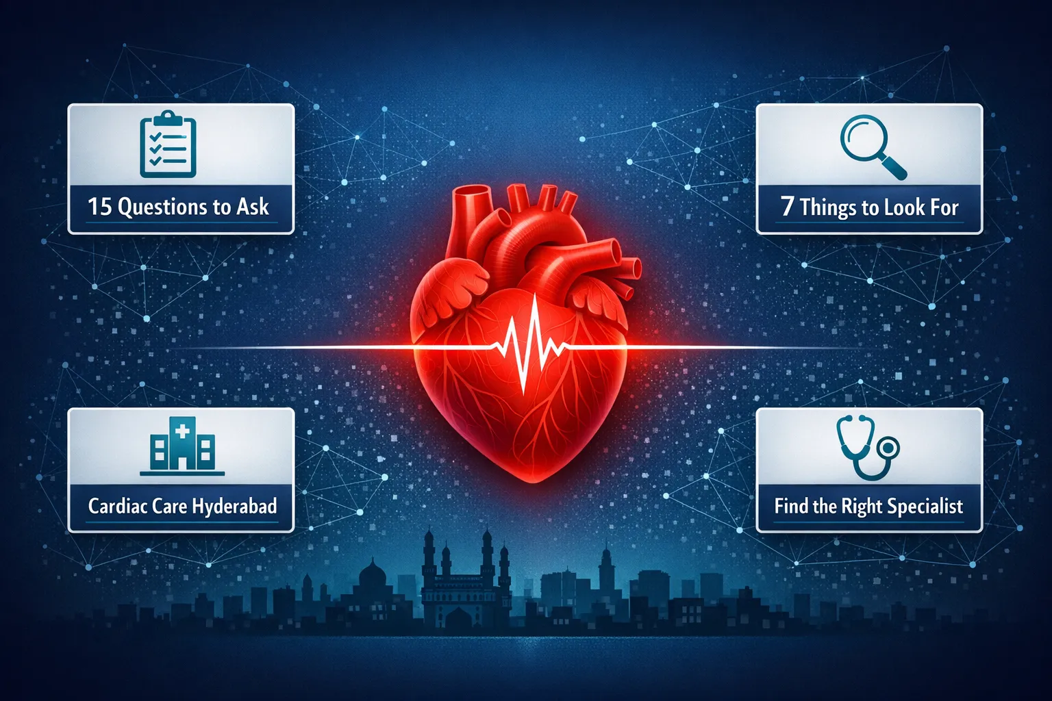
Find the best cardiologist in Hyderabad. Know what to look for, red flags to avoid, and 15 must-ask questions before your first cardiac specialist visit
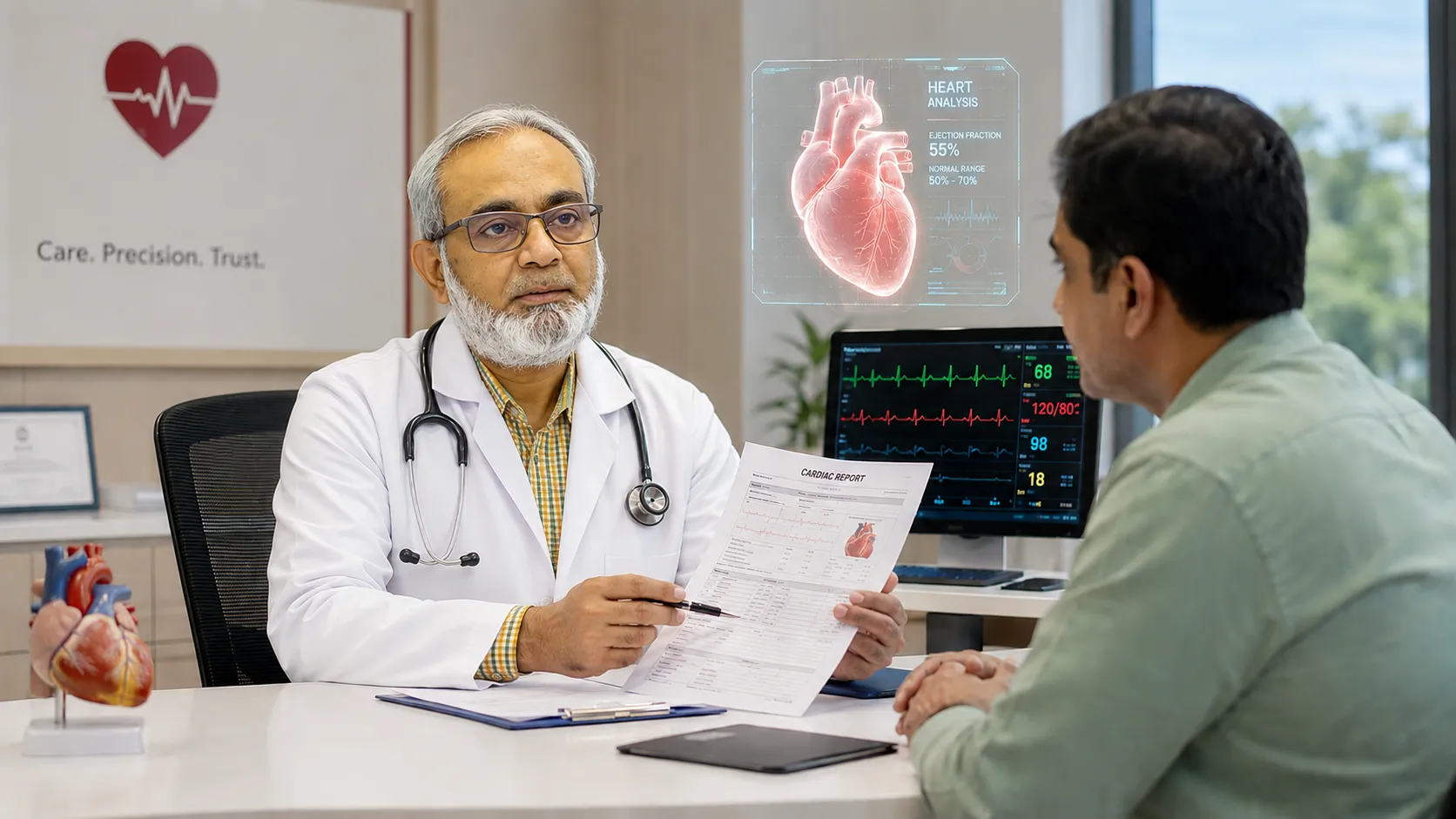
Read MoreLearn how a cardiologist supports better heart health through diagnosis, prevention, regular check-ups, lifestyle guidance and cardiac care in Hyderabad
Read MoreLooking for the best cardiologist in Attapur Hyderabad? Learn what to look for, key credentials to check, and why Dr Mohammed Wasif Azam Germanten at Germanten Hospital is a trusted choice for heart care
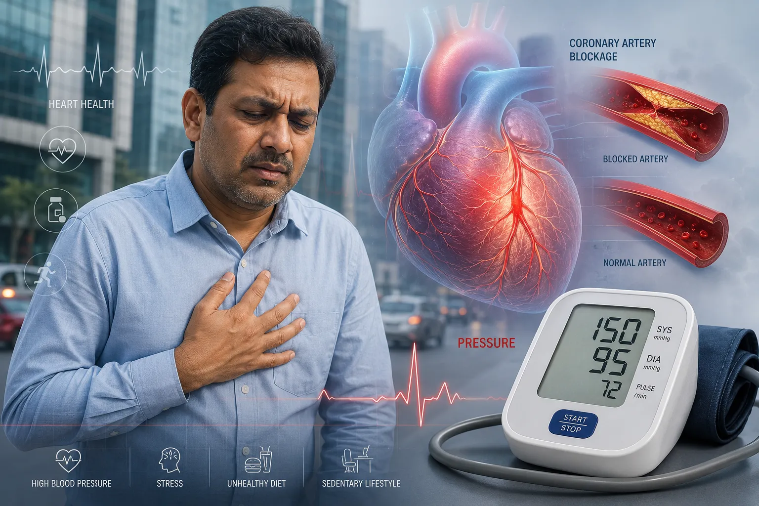
Read MoreUnderstand how high blood pressure silently damages your heart. A Hyderabad cardiologist explains the link between hypertension and heart disease, risk factors, and treatment options for patients in Attapur and across Hyderabad.
Read MoreDiscover the 10 most common heart disease risk factors in Hyderabad and how to reduce them. Expert advice from a preventive cardiologist in Attapur on diet, lifestyle, and cardiovascular prevention.
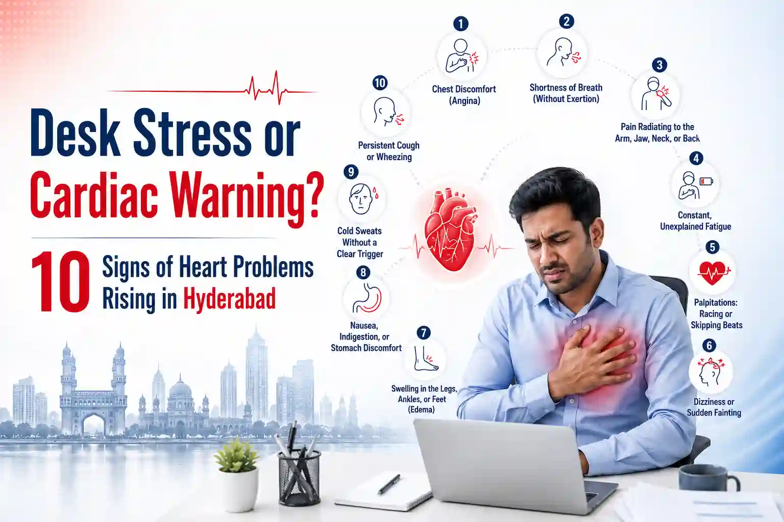
Read MoreExperiencing chest tightness or desk fatigue? Learn the 10 critical heart problem symptoms corporate workers in Hyderabad ignore. Book a checkup today.
Read MoreSpot the early signs of heart disease early, from chest pain to silent symptoms. Get expert cardiology care at Germanten Hospitals, Attapur, Hyderabad.
Read MoreGermanten Hospital is located in Attapur, one of South Hyderabad's most accessible neighborhoods. Patients from across southern and western Hyderabad can reach us within 20 to 30 minutes.